Peroxisome Under Electron Microscope
If you are looking for Human Biology Online Lab / Peroxisome- lab 2- Kaela Borders you've came to the right place. We have 14 Images about Human Biology Online Lab / Peroxisome- lab 2- Kaela Borders like Unique Characteristics of Eukaryotic Cells | Microbiology; Peroxisome Structure and Function ~ Biology Exams 4 U and also Human Biology Online Lab / Peroxisome- lab 2- Kaela Borders. Here you go:
Human Biology Online Lab / Peroxisome- Lab 2- Kaela Borders
 humanbiologylab.pbworks.com
 peroxisome lab kaela borders
Simultaneous Visualization Of Peroxisomes And Cytoskeletal Elements
 www.plantphysiol.org
 visualization actin peroxisomes cytoskeletal peroxisome simultaneous microtubule motility reveals elements plants based plantphysiol
Cytoplasma Dr.Jastrows EM-Atlas
 www.uni-mainz.de
 cytoplasma glatte muskulatur zytoplasma querschnitt filamen wartenberg externes wai
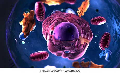
Peroxisomes Cell 3d Model
 www.turbosquid.com
 peroxisomes
Peroxisome Images; Stock Photos & Vectors | Shutterstock
 www.shutterstock.com
 peroxisome
Cytology: Definition And Basics | Online Medical Library
 www.lecturio.com
 peroxisome peroxisomes structure cell diagram organelles functions definition membrane cytology basics cytoplasm license cc organelle plant function lipid philschatz schatz
Squilla Mantis-Classification
 bioweb.uwlax.edu
 cell animal electron microscope plant cells under mantis shape anatomy alike terms squilla classification eukarya domain animals light differences
Unique Characteristics Of Eukaryotic Cells | Microbiology
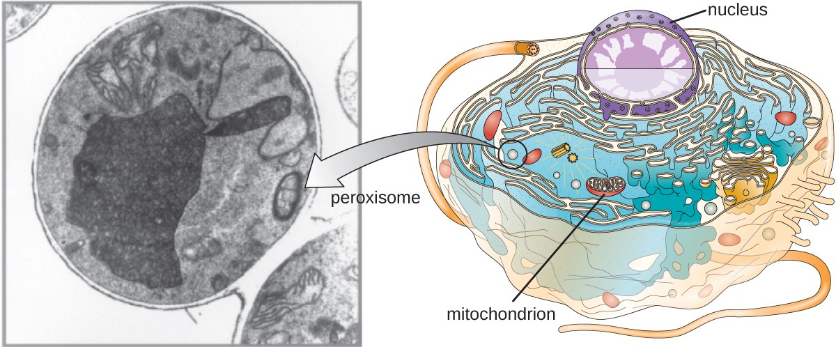 courses.lumenlearning.com
 peroxisome lysosome difference between cell peroxisomes eukaryotic cells characteristics structure function diagram location unique micrograph cytoskeleton electron containing figure libretexts
Electron Micrographs Of Cells Infected With Potato Virus Y (PVY). A
 www.researchgate.net
 electron pvy micrographs epidermal cytoplasm tissues organs vacuole
The Return Of The Peroxisome | Journal Of Cell Science
 jcs.biologists.org
 peroxisome
Peroxisome Structure And Function ~ Biology Exams 4 U
 www.biologyexams4u.com
 peroxisomes peroxisome structure function biology exams
Microbodies
 plantcellbiology.masters.grkraj.org
 structure peroxisome plant membrane enveloped structural contains wikipedia basic feature crystal unit inside single
De Histology: Cytoplasmic Deposits
 dehistology.blogspot.com
 electron glycogen micrograph histology figure cell deposits liver section
Euchromatin Dr.Jastrows EM-Atlas
 www.uni-mainz.de
 nucleolus euchromatin zelle zellkern golgi apparat heterochromatin electron nucleus microscopic organelles kern prolactin chromatin ratte
Electron glycogen micrograph histology figure cell deposits liver section. Cytology: definition and basics. Euchromatin dr.jastrows em-atlas
Posting Komentar untuk "Peroxisome Under Electron Microscope"